
100 consecutive cases of papillary cancer - case 002 (ultrasonographic picture 1)

|
|
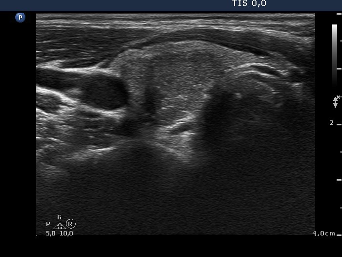
Right lobe, transverse scan. There is a hypoechogenic nodule presenting intranodular hyperechogenic figures of different types and a lobulated surface.