
100 consecutive cases of papillary cancer - case 002 (ultrasonographic picture 6)
|
|

|
|
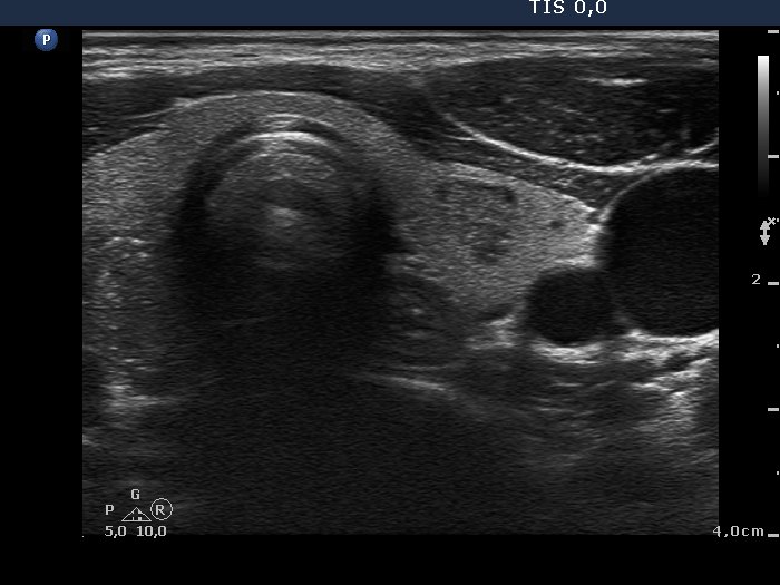
Left lobe, transverse view. There is a small, moderately hypoechogenic nodule in the central part of the lobe. It is worth comparing the non-specific intranodular granulations of the benign lesion with the brightest granules of the papillary carcinoma. A small part of the cancer can be seen left in the image.