100 consecutive cases of papillary cancer - case 026 (ultrasonographic picture 3)
|
|

|
|
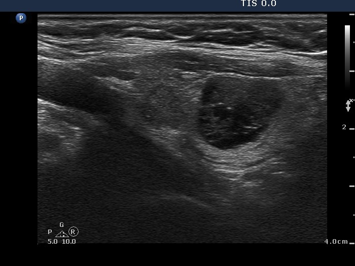
Right lobe, longitudinal scan. The mixed nodule presents back wall cystic figures.

2022-23 Advanced
Papillon Course
Supplementum
|
|
|
|
Right lobe, longitudinal scan. The mixed nodule presents back wall cystic figures.