
100 consecutive cases of papillary cancer - case 030 (ultrasonographic picture 10)
|
|

|
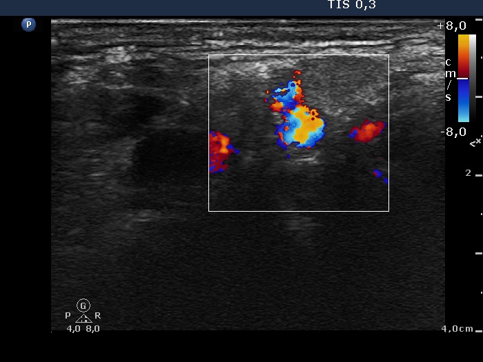
Left submandibular area of the neck, transverse view, color Doppler mode. The avascular hypoechogenic lesion left to the vessels proved to be a lymph node. Aspiration cytology disclosed a metastatic focus.