
100 consecutive cases of papillary cancer - case 032 (ultrasonographic picture 5)
|
|

|
|
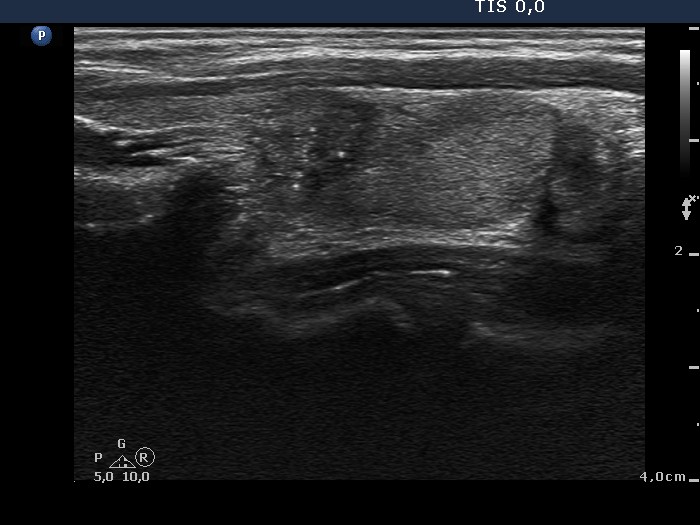
Middle third of the left lobe, another longitudinal scan. The nodule presents microcalcifications and lobulated margins.