
100 consecutive cases of papillary cancer - case 041 (ultrasonographic picture 5)
|
|

|
|
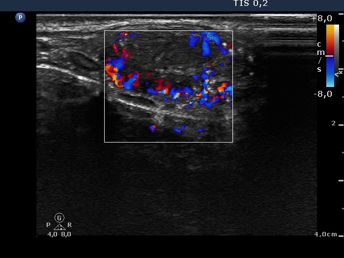
Left lobe, longitudinal scan, color Doppler mode. This image reveals a combined vascular pattern, the lesion has both perinodular and intranodular blood flow.