
|
|
100 consecutive cases of papillary cancer - case 056
|
|


Clinical presentation: A 50-year-old woman was referred for evaluation of mild hyperthyroidism. She was investigated of fatigue and 5 kg weight loss.
Palpation: Both lobes moderately firm. There was a nodule in the right lobe.
Functional state: euthyroidism (TSH 0.87 mIU/L, anti-TPO 665 U/mL).
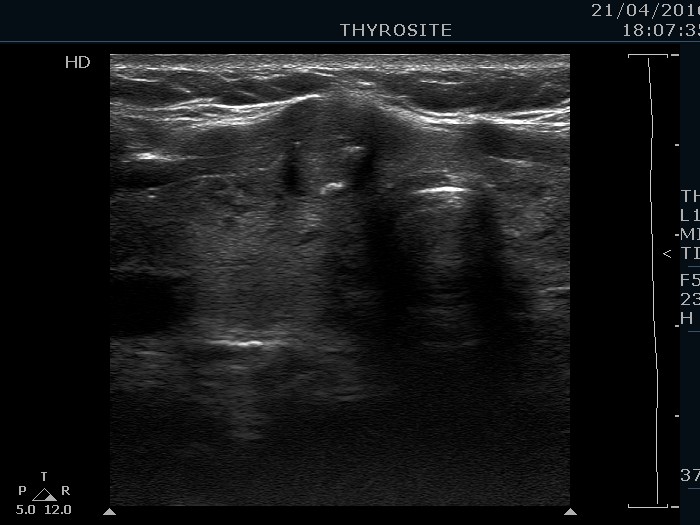
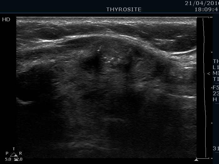
Ultrasonography. The thyroid was echonormal and contained hypoechogenic areas. The echogenicity index was around 20%. There was a moderately hypoechogenic lesion presenting microcalcifications in the ventral part of the right lobe.
Cytology resulted in papillary carcinoma.
Final diagnoses: Hashimoto's thyroiditis. Papillary carcinoma.
A total thyroidectomy was performed. Histopathology disclosed papillary carcinoma in the right lobe and chronic lymphocytic thyroiditis in the non-nodular part of the thyroid.