
|
|
100 consecutive cases of papillary cancer - case 069
|
|


Clinical presentation: A 49-year-old woman was referred for aspiration cytology. She has been treated for hyperthyroidism for 18 months. The patient observed a continuous increase in the left nodule over the past months.
Palpation: There was a large, firm nodule in the left lobe.
Laboratory tests: TSH 0.01 mIU/L, FT4 7.59 on daily 20 mg methimazole.
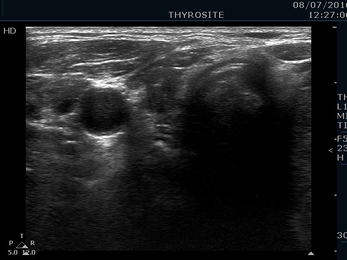
Ultrasonography. The thyroid was echonormal. There was a hypoechoic nodule in the right lobe. The nodule presented all three signs of a possible extrathyroidal extension. A large, mixed nodule occupied almost the entire left lobe. This lesion had back wall cystic figures and microcalcifications, too. The intranodular vascularization was increased.
US-guided aspiration was performed from both nodules. The cytology resulted in papillary carcinoma and in non-diagnostic report, right and left nodule, respectively.
Histopathology disclosed papillary carcinoma in the right nodule and follicular adenoma in the left nodule.
Comment.
- Such large nodules are difficult to examine and difficult to present in images.
- The left lobe clearly presented microcalcifications in the solid part.