
|
|
100 consecutive cases of papillary cancer - case 076
|
|


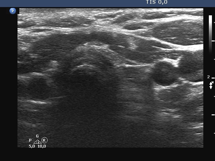


First examination (first two rows of images):
Clinical presentation: A 39-year-old woman went with her father to my consulting and she decided to request a thyroid examination. She has been treated with lithium for depression for more than 10 years. Two years after the initiation of antidepressant treatment hypothyroidism has developed. The patient was stopping the daily 50 microgram levothyroxine therapy one year ago because she felt no effect of the replacement treatment. Except for regular TSH determinations no other thyroid tests has been performed ever.
Palpation: both lobes were firm. There was a firmer nodule in the lateral part of the left lobe.
Laboratory tests: TSH 4.07 mIU/L, FT4 11.2 pM/L, aTPO 206 U/mL on daily 900 mg lithium.
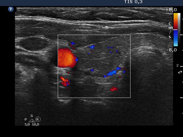
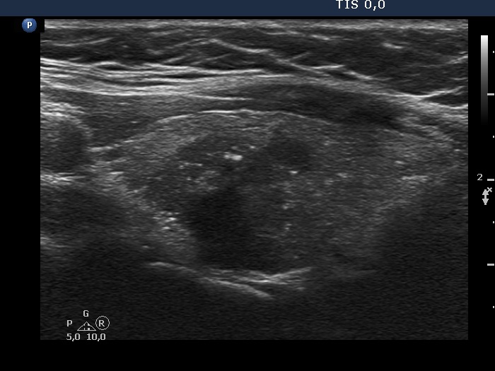
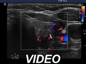
Ultrasonography. The thyroid was minimally-moderately hypoechoic and presented pronounced fibrotic changes. The left lobe had several hypoechoic nodules having cystic areas and numerous microcalcifications. Isolated microcalcifications were found in large numbers outside the nodules. The tumor showed irregularly increased intranodular vascularization.
Cytology. Papillary cancer.
Histopathology. Multifocal papillary cancer in the left lobe. The diameter of the largest focus was 20 mm. There were numerous smaller tumor foci, less than 2 mm in diameter.
Comments.
The ultrasound presentation is suspicious of invasive spread.
It is worth comparing the presentations of fibrosis in the right lobe and that of the microcalcifications outside the nodule in the left lobe.
Second examination a year after surgery (third row of images):
Clinical presentation: The patient underwent on radioidodine therapy. She had no complaints.
Palpation: no abnormality.
Laboratory tests: TSH 0.04 mIU/L, FT4 22.2 pM/L on daily 150 microgram levothyroxine. Thyroglobulin was 1.09 ng/mL, anti-hTg was below 0.9 U/mL.
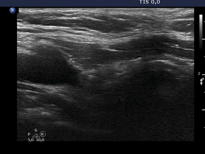
Ultrasonography. Both thyroid beds were filled with connective tissue. The left thyroid bed has a moderately hypoechoic area. Longitudinal section proved that this could not be a true nodule.