
100 consecutive cases of papillary cancer - case 077 (ultrasonographic picture 7)

|

|

|
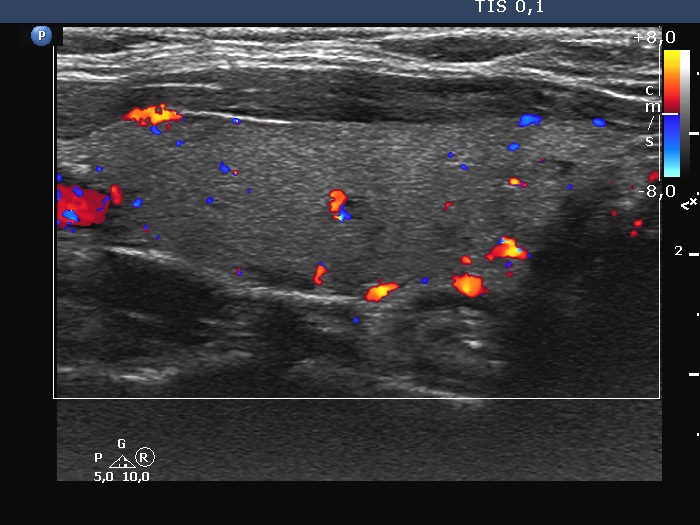
Left lobe, longitudinal scan, color Doppler mode. The hypoechoic, cystic tiny areas proved to be vessels.


