
100 consecutive cases of papillary cancer - case 080 (ultrasonographic picture 6)
|
|

|
|
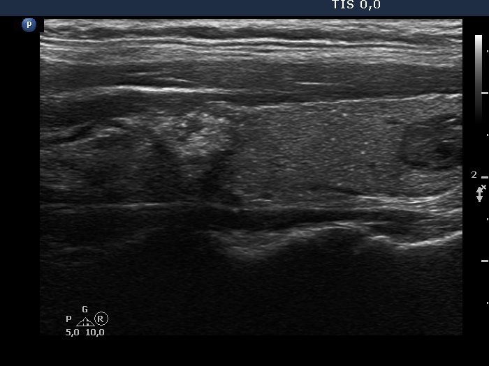
Left lobe, another longitudinal scan. Note the bright echogenic foci (microcalcifications) betweeen the two nodules.