
100 consecutive cases of papillary cancer - case 080 (ultrasonographic picture 10)
|
|

|
|
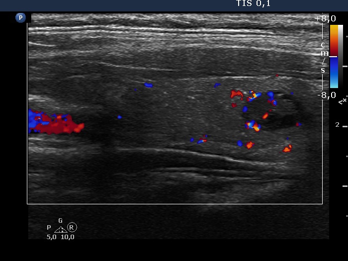
Lower part of the left lobe, longitudinal scan, color Doppler mode. The hypoechoic nodule has both intranodular vascularization and shows signs of perinodular blood flow, as well.