
100 consecutive cases of papillary cancer - case 081 (ultrasonographic picture 2)
|
|

|
|
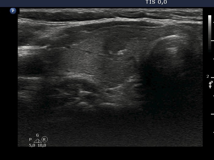
Right lobe, longitudinal. The more hypoechoic part of the lesion has a conglomerate of echogenic figures which are very suspicious of microcalcifications.