
100 consecutive cases of papillary cancer - case 086 (ultrasonographic picture 2)
|
|

|
|
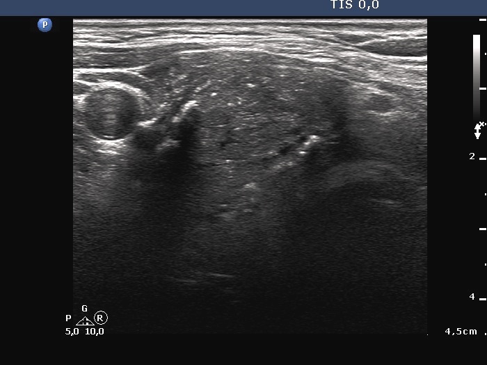
Right lobe, another transverse scan. The nodule has microcalcifications, macrocalcification and non-specific echogenic figures. The borders of the nodule cannot be clearly judged.