
100 consecutive cases of papillary cancer - case 089 (ultrasonographic picture 3)
Follow-up examination a year later
|
|

|
|
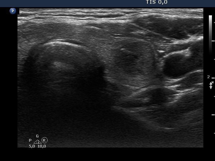
Left lobe, transverse scan. There is a hypoechoic nodule which has lobulated margins.