
|
|
100 consecutive cases of papillary cancer - case 097
|

|


Clinical presentation: A 17-year-old woman was referred for aspiration cytology of a thyroid nodule which was discovered on evaluation of irregular menstruation.
Palpation: a firm nodule in the isthmus.
Laboratory test: TSH 2.51 mIU/L, FT4 16.9 pM/L, aTPO 41 U/mL, ahTG 3.8 U/mL, TSAb undetectable.
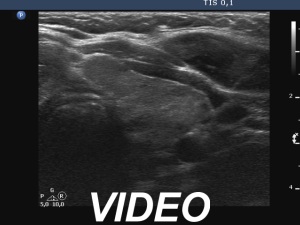
Ultrasonography. The thyroid was echonormal and had several hypoechoic areas. There was a hypoechoic nodule in the isthmus. The nodule had microcalcifications and back wall cystic figures. The borders were irregular. The lesion had scanty irregular vascularity.
Cytology resulted in papillary cancer.
Total thyroidectomy was performed. Histopathology disclosed a T2N1a papillary cancer. Beside the isthmic nodule, there was another papillary cancer focus in the left lobe which was 2 mm large. The extranodular thyroid tissue presented Hashimoto's thyroiditis. 10 out of th1 17 removed lymph nodes had metastatic focus.


