
|
|
100 consecutive cases of papillary cancer - case 100
|
|


Clinical data: A 38-year-old woman came to a follow-up. First, I met her 4 years ago when she was referred for evaluation of a newly diagnosed hypothyroidism at the sixth week of pregnancy. At that time a hypoechoic nodule was detected with the dimensions of 7, 8, and 9 mm, width, depth, and length, respectively. Replacement therapy was started and I suggested repeat ultrasound in a year.
Palpation: no abnormality.
Laboratory tests: 2.09 mIU/l on daily 50 microgram levothyroxine; aTPO level was 807 U/mL four years ago.
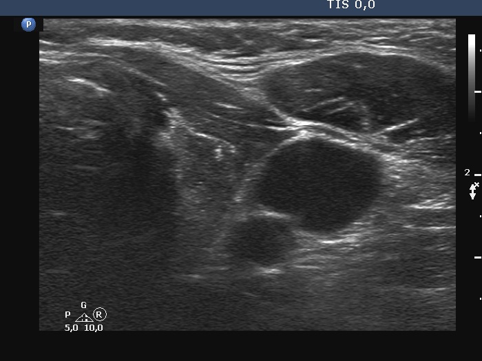
Ultrasonography. The thyroid was minimally hypoechoic. There were two discrete lesions in the left lobe. The larger has microcalcifications and irregular borders. The dimensions of this nodule were 9x11x12 mm (width, depth, length, respectively). The nodule had scanty intranodular vascularity.
Cytology resulted in papillary cancer.
A left lobectomy and left neck dissection were performed. Histopathology disclosed a T1b papillary cancer. No metastatic lymph nodes were found.