
|
|
The borders of the nodule - case 2024doi: 10.24390/thyrocase2024bord.00
|
|


Clinical presentation: A 34-yr-old woman was referred for follow-up examination at the 25th gestational week. She has been diagnosed with a nodular goiter for 8 years when she had a subacute granulomatous thyroiditis. At that time cytology resulted in benign colloid goiter. Actually, she had no complaints.
Palpation: no abnormality.
Laboratory test: TSH 0.97 mIU/L.
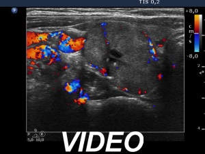
Ultrasonography. The thyroid was echonormal. There was an isoechoic mass in the right lobe. This mass was composed of multiple discrete lesions, which made the appearance of the entire lesion undulated, similarly to a cluster of grapes.
Cytology resulted in follicular tumor.
Right lobectomy was performed, and histopathology disclosed benign, hyperplastic nodules.
Comments.
-
The ultrasound presentation stands for hyperplastic nodules because there were multiple nodules next to each other.
-
This a typical example of non-pathological lobulation.