
The borders of the nodule - case 2157 (ultrasonographic picture 3)
doi: 10.24390/thyrocase2157bord.us03
|
|

|
|
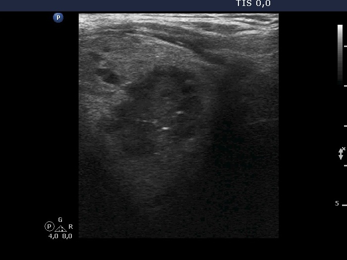
Right lobe, longitudinal scan. The hypoechoic part of the nodule has non-specific hyperechoic granules and two punctate hyperechoic foci (microcalcifications).