
The borders of the nodule - case conp 027 (ultrasonographic picture 5)
|
|

|
|
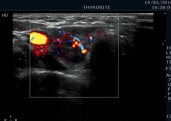
Lower part of the right lobe, transverse view, color Doppler mode. This is an irregularly increased, suspicious vascular pattern.