The borders of the nodule - case conp 037 (ultrasonographic picture 3)
|
|

|
|
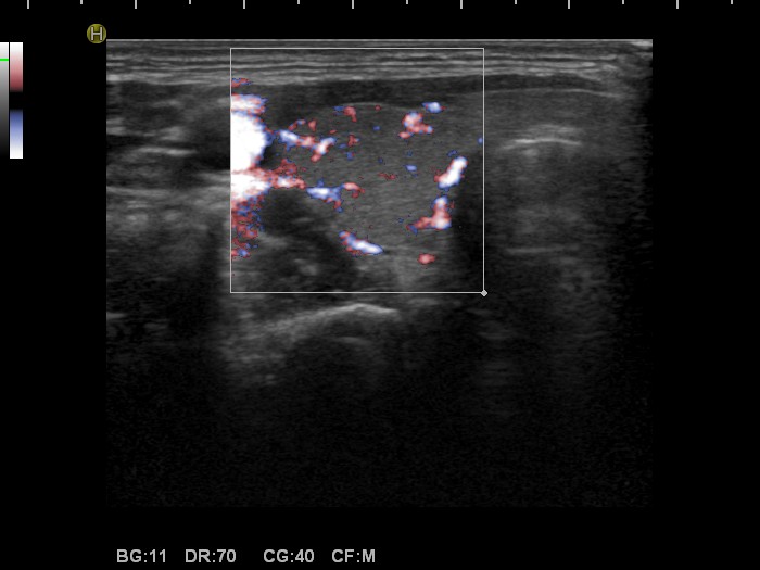
Right lobe, transverse view, color Doppler fine flow method. The vascularization is a little bit increased.

Nodule' borders
|
|
|
|
Right lobe, transverse view, color Doppler fine flow method. The vascularization is a little bit increased.