
|
|
The composition of the nodule - case 126
|
|

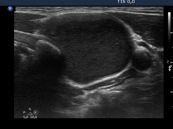


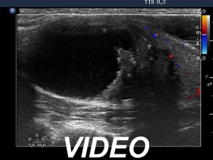

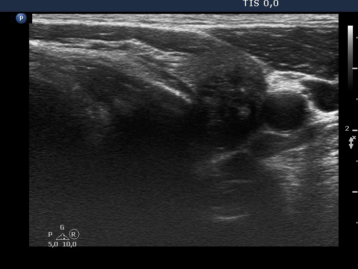
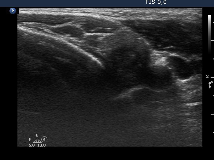

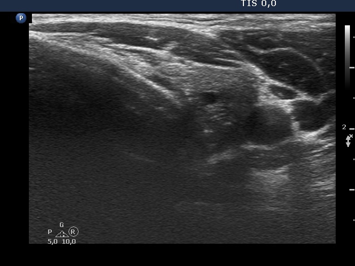
First examination (first and second rows of images):
Clinical presentation. A 43-year-old man was referred for ethanol sclerotherapy. He was diagnosed with a thyroid nodule two years ago. The nodule proved to be a cyst. The cyst has been evacuated three-times, but it has been always refilled within weeks. The nodule caused difficulties in swallowing.
Palpation: a firm nodule in the left lobe.
Hormonal evaluation: euthyroidism with TSH 0.90 mIU/L.
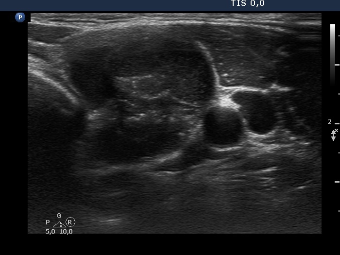
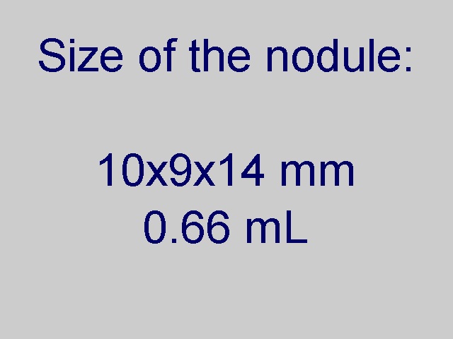
Ultrasonography. The thyroid was echonormal. The left lobe had a large nodule. The upper 4/5 of the lesion was hypoechogenic but not anechoic. There was a minimally hypoechogenic solid part surrounded with multiple small cystic chambers in the lower 1/5 of the nodule. The solid area had numerous echogenic figures.
We removed 13 mL brown fluid and thereafter injected 3 mL ethanol.
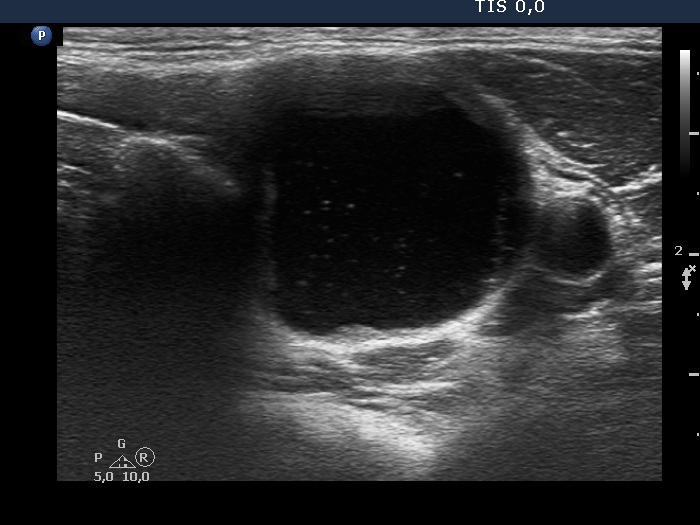
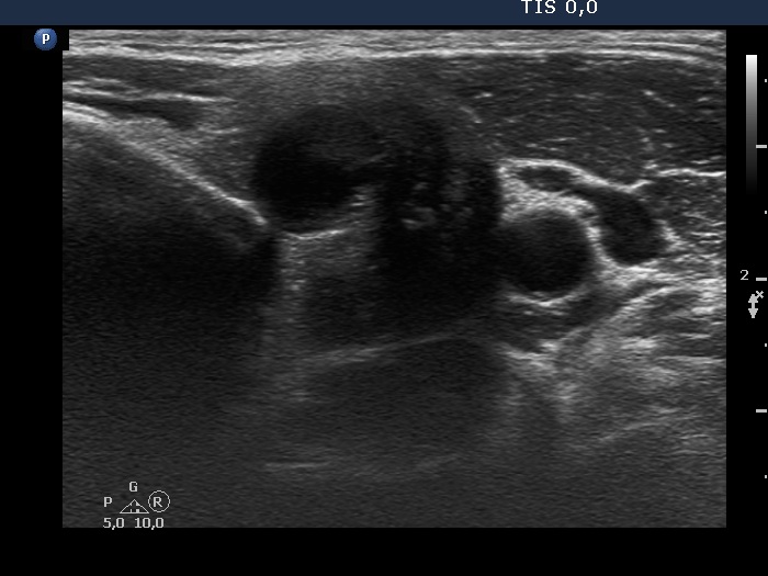
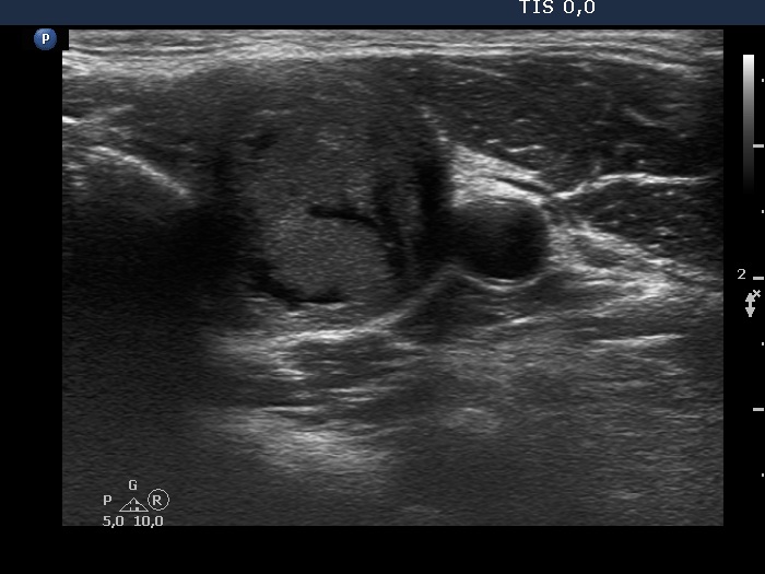
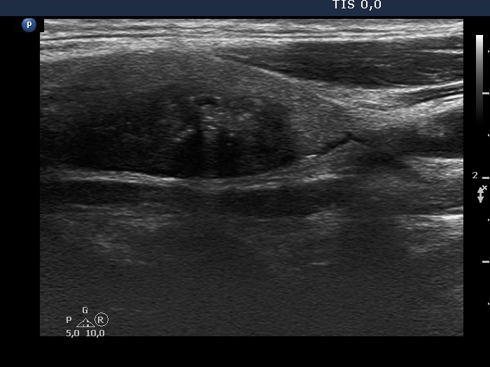
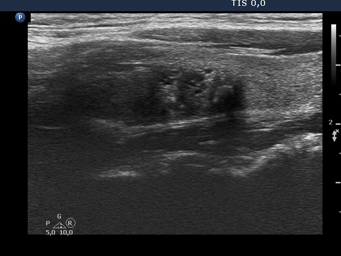
Second and fifth sessions of sclerotherapy (second and third rows of images, respectively)
Clinical presentation. Five sessions of ethanol sclerotherapy were administered. At the second attempt 10 mL brown fluid was aspirated and 1.1 mL ethanol was injected. These amounts were 7 mL and 4 mL, 1 mL and 1.5 mL, removed cystic fluid and injected ethanol, third and fourth session, respectively.
We could aspirate 2 ml thick, brown cystic fluid and injected 3 mL ethanol.
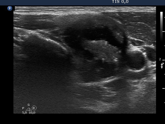
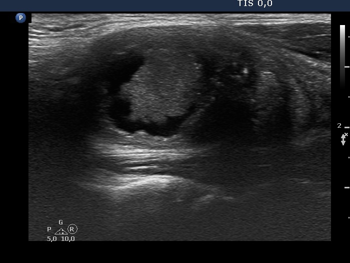
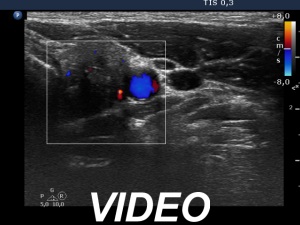
Follow-up investigations 6 weeks after the last session (fourth row of images)
Clinical presentation. The patient did not feel the nodule. The difficulties in swallowing have ceased.
Ultrasonography. The lesion underwent dramatic shrinkage.
Follow-up investigations 6 months after the last session (fifth row of images)
Clinical presentation. The patient had no complaints.
Ultrasonography. A further significant decrease was detected.
Comments.
- This is a rare but not exceptional situation that the pattern of a cyst is not anechoic but only deeply hypoechogenic. It is reasonable to assume that such nodules are filled with thick, protein-rich fluid or more likely they contain elements of blood.
- The echo pattern of a nodule underwent on sclerotherapy can turn to a suspicious pattern. This normal phenomenon includes the appearance of various echogenic figures mimicking microcalcifications, lobulated shape of a solid part undergoing on shrinkage. This deceptive presentation might cause even great concern if the ultrasonographer is not aware either of the previous sclerotherapy or of the presentation of a sclerotized nodule. Therefore, we always pay the attention of our patients after the sclerotherapy that they should not be afraid if on thyroid ultrasound examination that is being performed later in their lifetime "suspicious" findings will be described.