
The composition of the nodule - case 1457 (ultrasonographic picture 8)
|
|

|
|
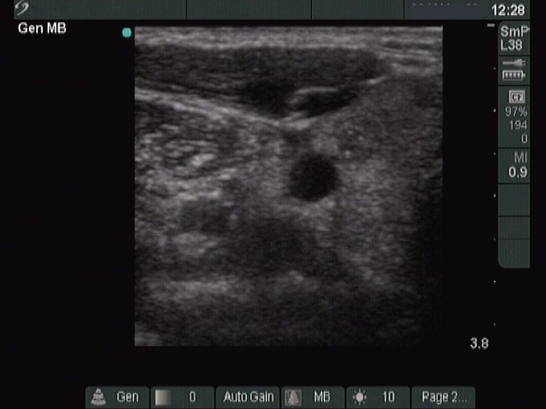
Right lobe, transverse view - after the aspiration of 0.4 mL cystic fluid. The presentation of the previously almost completely cystic lesion has substantially changed. Before the aspiration, it was ambiguous whether the lesion had solid parts, while after the removal of the cystic fluid, a moderately hypoechogenic solid part has appeared which contains several punctate echogenic foci, microcalcifications.