
|
|
The composition of the nodule - case 491
|
|


First examination (first row of images):
Clinical presentation: A 65-year-old woman was referred for evaluation of a recurrent nodule. She has undergone right subtotal lobectomy for 15 years. She noticed a lump in the left lobe six month before the present examination.
Palpation: The left lobe had a firm nodule.
Result of blood test: TSH 1.58 mIU/L.
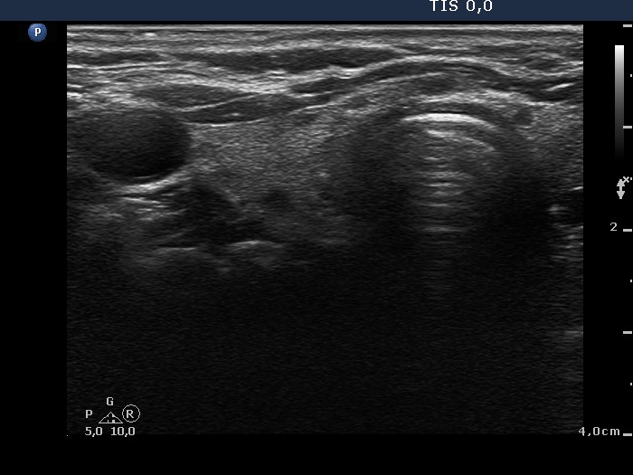
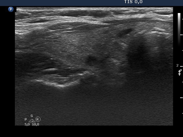
Ultrasonography. The right lobe was echonormal and had several hypoechogenic areas. The left lobe had a large cystic nodule which belonged to the central subtype. The lesion had echogenic figures caused by back wall posterior enhancement. The dimensions of the nodule were 31, 21 and 40 mm (width, depth and length, respectively).1.5 mL serous fluid was aspirated. Aspiration cytology resulted in benign cystic lesion.
We suggested follow-up because the nodule did not cause any problem to the patient.
Second examination 3 years later (second and third rows of images):
Clinical presentation: The patient came to routine follow-up visit. She had no complaints.
Palpation: The left lobe had a firm nodule.
Result of blood test: TSH 1.03 mIU/L.
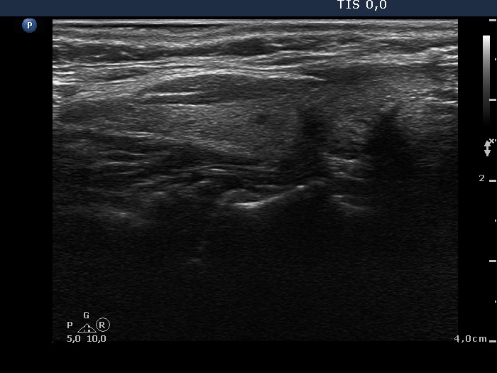
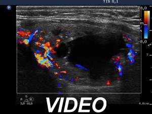
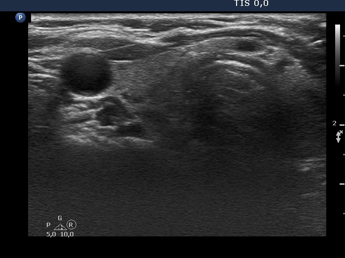
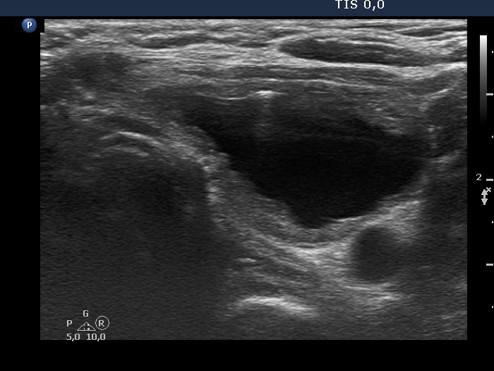
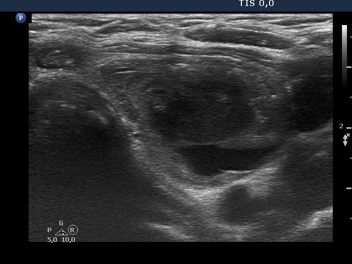
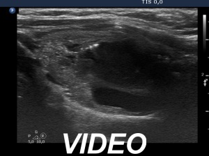
Ultrasonography. The presentation of the nodule remained unchanged. The dimensions of the nodule were 33, 18 and 41 mm (width, depth and length, respectively).We aspirated 5 mL bloody cystic fluid. At the end of the aspiration we noticed that cyst started to refill at once. 30 seconds after finishing the aspiration, the cyst had regrown to the previous size. (See video record.)
Suggestion: repeat examination in 6 months, in the event of complaints at once.