
|
|
The composition of the nodule - case conp 050
|
|


Clinical presentation: A 43-year-old man was referred for aspiration cytology. He discovered a lump in the right side of the neck several month ago.
Palpation: There was a firm lump lateral to the right thyroid. The right lobe was nodular on palpation.
Laboratory tests: euthyroidism.
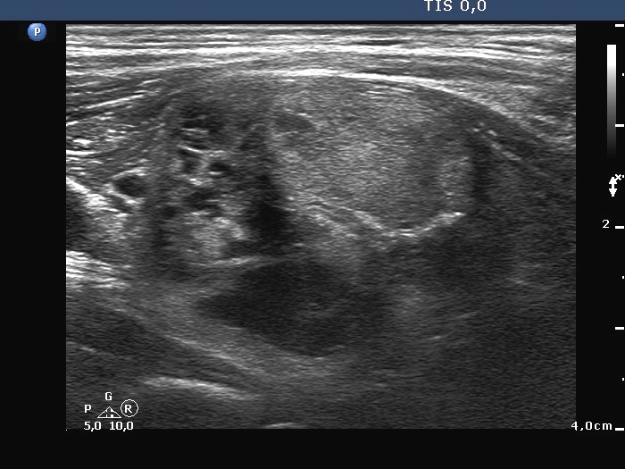
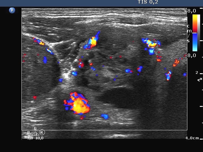
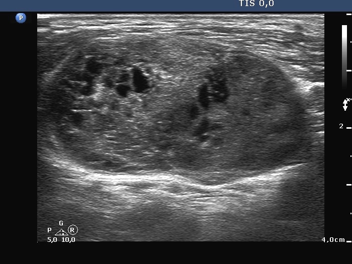
Ultrasonography. The right lobe contained multiple nodules presenting cystic degeneration. The largest nodule was echonormal. The left thyroid was intact. According to the palpable mass there was a distinct lesion lateral to the right carotid artery. This mass was composed of multiple discrete cystic areas. However, only around half of the mass was composed of tiny cystic areas, therefore the pattern did not meet the criteria of a spongiform cyst. The vascularization of the right lobe and the aberrant thyroid tissue was non-specific and scanty.
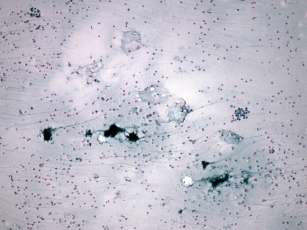
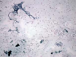
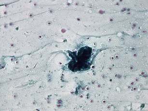
Cytology was performed from the aberrant tissue and the right thyroid. On all three occasions a small amount of serous material was gained. The cytological patterns were identical. We demonstrate here, the images of the smear gained form the aberrant thyroid. Cytological diagnosis was benign cystic lesion.
A right lobectomy and the removal of the lateral mass was performed. Histopathology disclosed benign hyperplastic nodules corresponding to the right lobe. The removed lateral mass was 5 cm in maximal diameter and contained hyperplastic nodules and a focus of a follicular variant of a papillary carcinoma without any signs of invasion. The maximal diameter of the tumor was 2.5 cm.
Comment.
-
The tumor originated in an aberrant thyroid tissue.
-
By reviewing the smear, we could not find any suspicious cytological signs. It means that the cause for false-negative result was the failure of the aspiration. Note that the volume-ratio of the carcinoma to the whole aberrant mass was around 1:8.
-
It is worth analysing the video record. There was a more solid area in the medial part of the mass. We suppose that the tumor was located in this part of the aberrant tissue while we aspirated the central part of the mass.
.