
Intranodular hyperechogenic figures - case 1139 (ultrasonographic picture 4b)
|
|

|
|
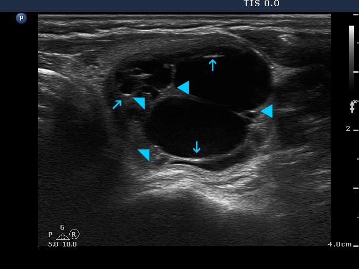
Left lobe, another transverse scan. The posterior acoustic enhancement in the dorsal wall of the small cystic areas causes no concern if this optical artifact is linear (arrows), however granular figures might be misinterpreted as microcalcifications (arrowheads).