
Intranodular hyperechogenic figures - case 1139 (ultrasonographic picture 8)
|
|

|
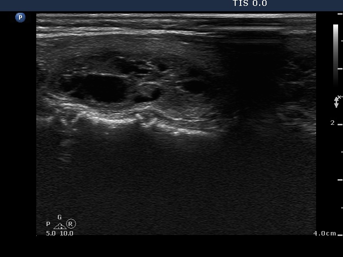
Left lobe, longitudinal scan - after aspirating 4.5 ml pale yellow fluid. The hyperechogenic figures are caused by back wall posterior enhancement.