
|
|
Intranodular hyperechogenic figures - case 1343
|
|


Clinical presentation: A 55-year-old woman was referred for neck ultrasound and aspiration cytology of a suspicious nodule described on ultrasound examination. The patient has noticed a painless hard lump for two months in the right side of the neck. The patient was operated on fifteen years ago when histopathology disclosed an oxyphilic variant of papillary carcinoma. Total thyroidectomy was performed and the patient received external irradiation because of the lack of radioiodine uptake. The thyroglobulin level became undetectable and remained so up to these days. The anti-hTg level varied between 38 and 210 U/mL without any tendency.
Palpation: a hard mass in the right thyroid bed.
Functional state: euthyroidism on daily 112.5 microgram levothyroxine with TSH 0.92 mIU/L. Thyroglobulin was undetectable, anti-hTg was 54 U/mL.
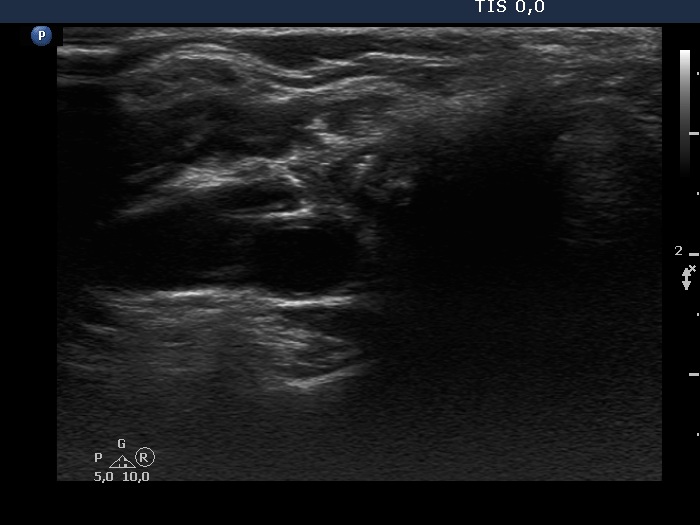
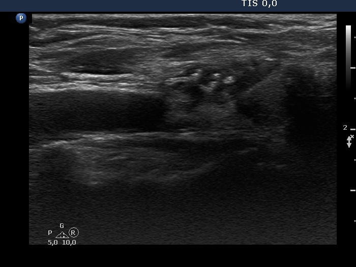
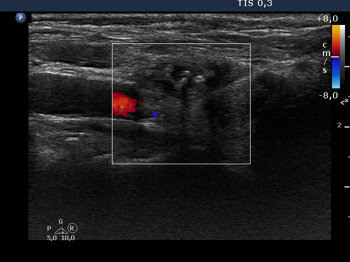
Ultrasonography. There was no thyroid parenchyma. A hypoechogenic lesion was found in the right thyroid bed. The lesion had an irregular shape, it contained ragged, moderately hyperechogenic tissue and relatively large, hyperechogenic figures. The lesion was avascular.
Cytology: granulation around surgical thread.
Histopathology: granulation around surgical thread.
Comment.
-
The presentation of a granulation around surgical thread is close to that of papillary carcinoma. On the other hand, the granules in the former are larger and in contrast with the malignant disease these figures are not round and are located in an echonormal patch.
-
A granulation around surgical thread might appear even decades after the surgery.