
|
|
Intranodular hyperechogenic figures - case 1365
|
|


Clinical data: A 41-year-old woman requested a second opinion. She was examined because of hypothyroidism and a newly discovered nodule in another outpatient department three months ago. Aspiration cytology resulted in atypia of an unknown significance. The diagnosis made the patient very anxious. The patient had great fear both from harboring thyroid carcinoma and from the surgical procedure, too. She has already got anti-depressants before visiting us.
Functional state: euthyroidism on daily 37.5 microgram levothyroxine.
Palpation: no abnormality.
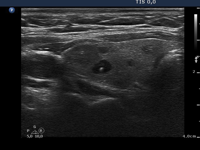
Ultrasonography: The thyroid was minimally hypoechogenic. There were several small cystic areas in both lobes and a moderately hypoechogenic, inhomogeneous lesion in the right lobe. The latter displayed no vascularity and was aspirated previously.
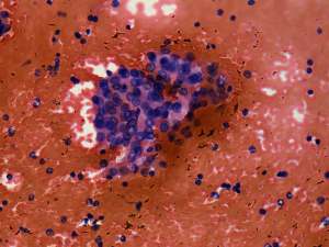
Aspiration cytology: There was no colloid in the background. Follicular cells occurred in microfollicles and compact clusters. Lymphocytes were also present in smaller number, even mixed with follicular cells. Grooves were found occasionally.
Combined sonographic-cytological diagnosis: Chronic lymphocytic thyroiditis. The risk of malignancy is not greater than 1%.
We advised follow-up examinations instead of surgery. However, the patient wanted to hear that malignancy can be excluded with 100% safety. Naturally, one cannot exclude malignancy 100%. She decided to undergo surgery.
Histopathology disclosed Hashimoto's thyroiditis with several cystic areas but without any nodule.
Comments:
-
The sonographic pattern imitated a nodular goiter; a frequent situation in Hashimoto's thyroiditis.
-
One of the greatest practical problem with category AUS of Bethesda system is demonstrated in this patient: facing with a risk of malignancy in the range of 10-20%, most patients decide to undergo surgery instead of follow-up examinations.
-
We prefer a combined sonographic-cytological diagnosis instead the Bethesda system. In this case the cytological picture was not calming. However, considering the sonographic pattern the risk of malignancy was very low.