
Intranodular hyperechogenic figures - case 1386
(ultrasonographic picture 1a)
|
|

|
|
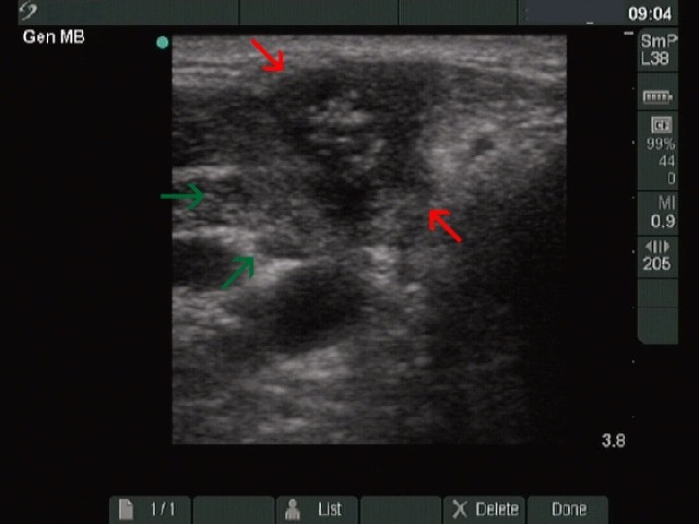
Right lobe transverse scan. There is a hypoechogenic inhomogeneous lesion marked with red ventral to the operated thyroid marked with green. The lesion contains a hyperechogenic patch with small bright hyperechogenic granules within it. This pattern is very similar to an amyloid deposit seen in medullary cancer.