
Intranodular hyperechogenic figures - case 1473 (ultrasonographic picture 7)
|
|

|
|
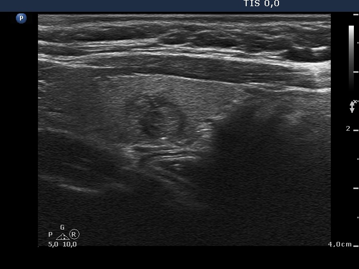
Left lobe, longitudinal view. The minimally hypoechogenic lesion presents halo sign. The bright echogenic figure in the dorsal surface of the nodule is solitary, a bit elongated and is related to a tiny ventral cystic area. These features stand for a back wall figure. Nevertheless, the possibility of a punctate echogenic focus (microcalcification) cannot be excluded.