
|
|
Intranodular hyperechogenic figures - case 1490 |
|


Clinical presentation: A 33-year-old woman was referred for aspiration cytology of a nodule detected on screening.
Palpation: no abnormality.
Functional state: euthyroidism with TSH 1.41 mIU/L.
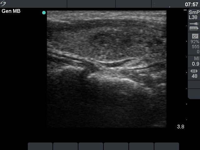
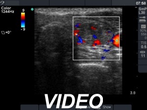
Ultrasonography. The thyroid was echonormal. There was a hypoechoic nodule in the left lobe. The lesion echogenic granules. Great proportion of these granules had a dorsal narrowing tail, therefore, they correspond to colloid crystals, comet-tail artifacts. The nodule presented signs of perinodular and intranodular blood flow.
Aspiration cytology was performed two times and resulted in non-diagnostic report. Only scattered number of follicular cells were found. Although thyrocytes did not show atypia, the number of cells and cell groups was not enough for a cytological diagnosis.
The patient wished to be operated on.
A left lobectomy was performed. Histopathology disclosed follicular adenoma.
Comments. It is worth analysing the video, at first sight the bright granules seem to be microcalcifications.