
|
|
Intranodular hyperechogenic figures - case 1518
|
|


Clinical presentation: A 74-year-old woman was referred for evaluation of a nodular goiter detected on carotid Doppler examination. The patient was investigated because of nephrotic syndrome.
Palpation: a not firm nodule in the left lobe.
Functional state: euthyroidism with TSH 3.19 mIU/L.
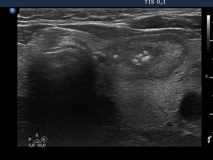
Ultrasonography. The thyroid was echonormal and contained several nodules of different echogenicities including a hypoechogenic one in the dorsal part of the left lobe and a minimally hypoechogenic lesion presenting groups of echogenic foci in the ventral part of the left lobe.
Aspiration cytology of the nodule in the right lobe resulted in benign follicular proliferation (Bethesda 3) while did in benign colloid goiter in the event of the left nodule.
Comments.
-
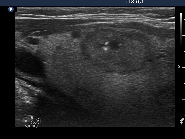
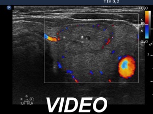
A single bright echogenic focus within the right nodule can be seen in the video. This figure belongs to punctate echogenic foci subgroup.
-
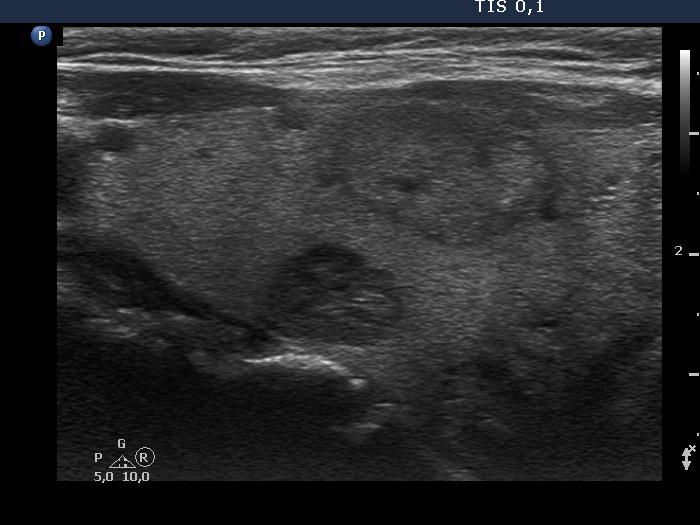
The origin of the echogenic foci within the left nodule is equivocal. They probably correspond to groups of smaller echogenic foci.