
|
|
Intranodular hyperechogenic figures - case 1539
|
|


Clinical presentation: A 35-year-old woman was referred for evaluation of a suspected multifocal papillary carcinoma detected on ultrasound examination of a manager screening. On the ultrasound report multiple hypoechogenic nodules containing numerous microcalcifications were described. The patient was told orally that she harbors a multifocal papillary carcinoma with great probability.
Palpation: no abnormality.
Functional state: euthyroidism with TSH 1.76 mIU/L.
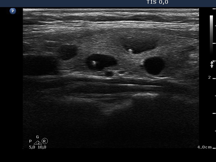
Ultrasonography. The thyroid was echonormal. There were numerous cystic areas in both lobes. They were less than 1 cm in maximal diameter and contained intranodular hyperechogenic granules. The latter were bright and except for two figures presented with the characteristic dorsal narrowing tail of comet-tail artifact.
Two of the cystic lesions were aspirated and cytology resulted in benign cystic-colloid lesion in both locations.
I tried to calm the very anxious patient and told her that the possibility of carcinoma can be excluded and even these lesions cannot be held as abnormal.
She visited another thyroidologist who did not perform ultrasound himself. He told the patient that the risk of carcinoma is minimal but if she cannot bear the existing uncertainty about the diagnosis it would be better to undergo surgery.
A right lobectomy was performed. An intact thyroid with several cystically dilated macrofollicles was described on histopathology.
Comments.
-
The first concern is the ultrasound screening which serves particular economic interest but does not serve the interest of the person whom this is proposed for. There are very rare exceptions, i.e. a known primary carcinoma, familiar history of thyroid carcinoma and previous irradiation of the thyroid area. Otherwise, the performance of thyroid ultrasound with no abnormality on palpation in a person not known suffering or has been suffered from any thyroid disorder is a very bad and harmful practice.
-
Except for very few cases, to raise malignancy on a thyroid ultrasound report is a serious failure. In this particular case, the professional issue was the misinterpretation of comet-tail artifacts as microcalcifications.
-
The presentation of comet-tail artifacts varies depending on the anatomical situation, the depth, the thickness of the fluid and on the ultrasound device. Occasionally, a comet-tail artifact is very similar to a microcalcification. On the other hand, by judging an actual hyperechogenic figure we have to take into account the ultrasound pattern of the whole nodule. There were two figures which lacked the typical dorsal narrowing tail but there were many other typical forms. So, these two questionable figures have to be held also as comet-tail artifacts.
-
The pivotal of the management of thyroid patients is to avoid causing unnecessary anxiety. If we behave inappropriately than it is very hard, occasionally - as happened in this case - impossible to reassure a patient. We are aware that a thyroid nodule extremely rarely causes any long-term decrease in life quality or life expectancy. A thyroidologist is aware about this fact but a patient isn't.