
Intranodular hyperechogenic figures - case 155
Three years after the previous examination (ultrasonographic picture 6)
|
|

|
|
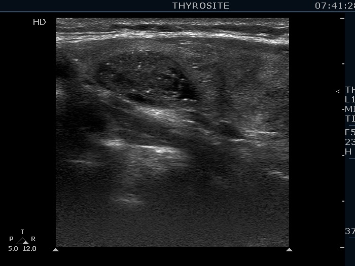
Left lobe, longitudinal scan. There are numerous hyperechogenic granules which might be microcalcifications on still image. On the other hand, the whole presentation of this lesion is benign, first of all because of the regular geometrical shape and the sharp borders. For papillary cancer, it would be unusual for the margins of the nodule to remain completely regular for such a large number of microcalcifications. The video proves that most of these bright granules are related to tiny ventral cystic areas, therefore these are presentations of back wall posterior enhancement.