
Intranodular hyperechogenic figures - case 1726 (ultrasonographic picture 2)
|
|

|
|
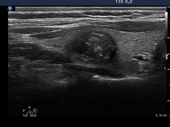
Right lobe, longitudinal view. Beside microcalcifications, there are less bright and smaller hyperechogenic granules and short lines within the nodule. The latter figures correspond to connective tissue.