
Intranodular hyperechogenic figures - case 1789 (ultrasonographic picture 4)
|
|

|
|
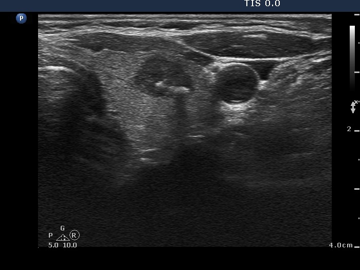
Left lobe, trasnverse view. There is a hypoechogenic nodule with sharp but lobulated margins. The lesion contains two hyperechogenic figures. The medial one (left in the image) is difficult to interpret while the lateral irregular thicker one (right in the image) is a coarse calcification on the presence of dorsal acoustic shadow.