
|
|
Intranodular hyperechogenic figures - case 2174
|
|


Clinical presentation: A 49-year-old woman was referred for evaluation of a nodular goiter detected on screening.
Palpation: no abnormality.
Functional state: euthyroidism with TSH 1.64 mIU/L.
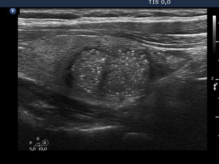
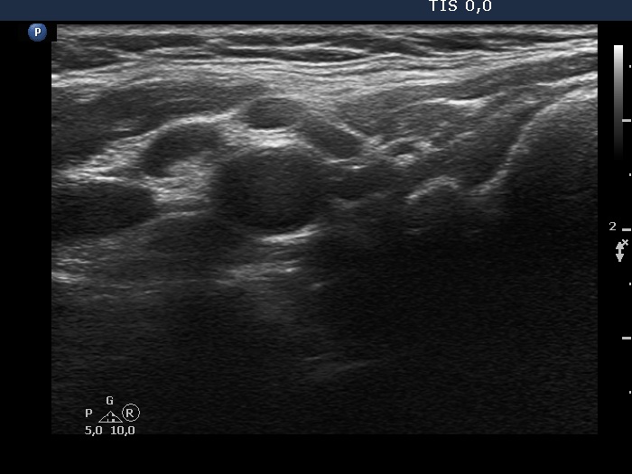
Ultrasonography. The thyroid was echonormal. There was a hypoechogenic nodule in the right lobe. The nodule had numerous punctate echogenic granules.
Three aspiration was performed. Cytology resulted in benign cystic-colloid lesion.
Suggestion repeat ultrasound and aspiration cytology in a year.
Comments.
-
The echogenic figures are very difficult not to interpret as microcalcifications. Nevertheless, thorough analysis reveals that some of the bright granules has a short tale, and great proportion of these figures are related to ventral tiny hypoechoic areas, probably cysts. Moreover, there are not only echogenic granules but also short echogenic lines within the nodule. Taking all in all, these echogenic figures are very suspicious being microcalcifications.
- The ACR TIRADS is the only one which does not use the term 'microcalcification', instead they use the term 'punctate echogenic foci', which includes microcalcifications and short-tail comet-tail artifact. This example stands for the advantage of the ACR terminology.
- In such nodules it is to be considered to delay the ultrasound report till the result of FNA. In this patient FNA disclosed benign colloid goiter and there were macrophages on the smear. The latter proved that the nodule had cystic areas. If we take this fact into account, than it can solve our concern about the interpretation of echogenic granules.