
Intranodular hyperechogenic figures - case 2174 (ultrasonographic picture 2b)
|
|

|
|
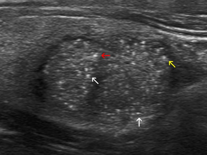
Right lobe, longitudinal view, enlargement. Some of the echogenic granules are located dorsal to more hypoechoic areas (white arrows), which raises the possibility of back wall cystic enhancement as a cause of these figures. At least one of the granules has a dorsal tail (yellow arrow). On the other hand, the punctate echogenic focus (marked with a red arrow) fulfills the criteria of microcalcification.