
|
|
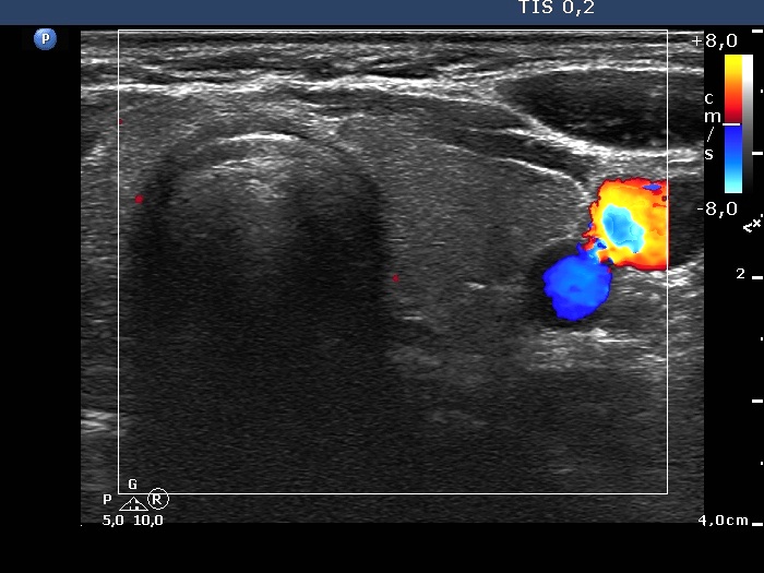
Intranodular hyperechogenic figures - case 28
|
|


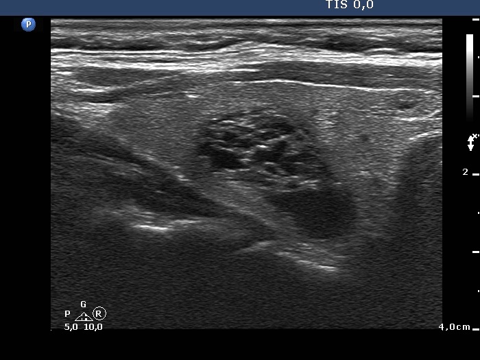


Clinical presentation: A 66-year-old woman was referred for follow-up of a nodule discovered on screening 6 years ago. The woman had no complaints.
Palpation: no abnormality.
Functional state: euthyroidism with TSH 1.72 mIU/L.
Ultrasonography. The thyroid was echonormal. There was a spongiform-type cyst in the right lobe. The small cystic areas of the lesion exhibited posterior back wall enhancement. The vascularization was scanty. Compared with the former examination the nodule remained just as large.
Suggestion: repeat ultrasound in five years.
Comments. Note the typical presentation of posterior back wall enhancement.