
|
|
Intranodular hyperechogenic figures - case 367
|
|


Clinical presentation: A 39-year-old woman was referred for aspiration cytology of a suddenly developed lump.
Palpation: an elastic nodule in the right lobe.
Functional state: euthyroidism with TSH 0.71 mIU/L.
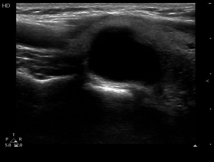
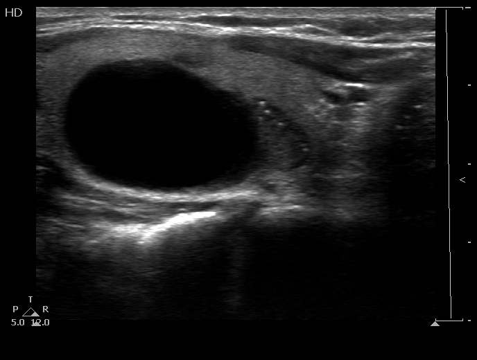
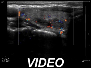
Ultrasonography. The thyroid was echonormal. There were two lesions in the right lobe, the upper was a calcified one while the lower was a cystic nodule. Next to the latter there was a hypoechogenic lesion presenting intranodular figures including microcalcifications. It was equivocal whether this hypoechogenic area was part of the cystic nodule or a distinct other lesion. After aspirating 6.5 mL brown fluid, it became easier to judge the hypoechogenic lesion, it had shaper borders and a regular geometrical shape and presented perilesional blood flow.
Cytology resulted in cystic degeneration.
We gave a combined ultrasound-cytological diagnosis of suspicion of papillary carcinoma.
Histopathology disclosed follicular adenoma.
Comment. The lesion seemed to be suspicious because of the presence of several intranodular hyperechogenic granules. On the other hand, the nodule was not irregularly shaped and had sharp borders. As the final histopathology proved, we overinterpreted the ultrasound presentation.