
|
|
Intranodular hyperechogenic figures - case 372
|
|


Clinical presentation: A 55-year-old woman has noticed a suddenly developed lump in her right lobe for 3 weeks. She was known harboring a cystic nodule for 3 years when 2.5 mL cystic fluid was aspirated.
Palpation: a not firm nodule in the right lobe.
Functional state: euthyroidism with TSH 1.07 mIU/L.
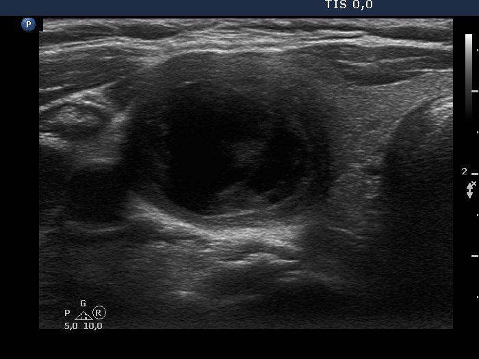
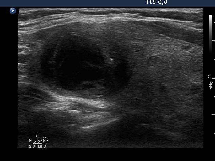
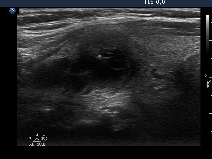
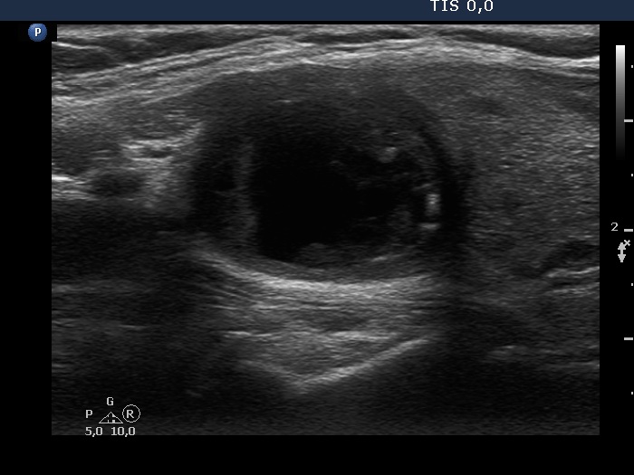
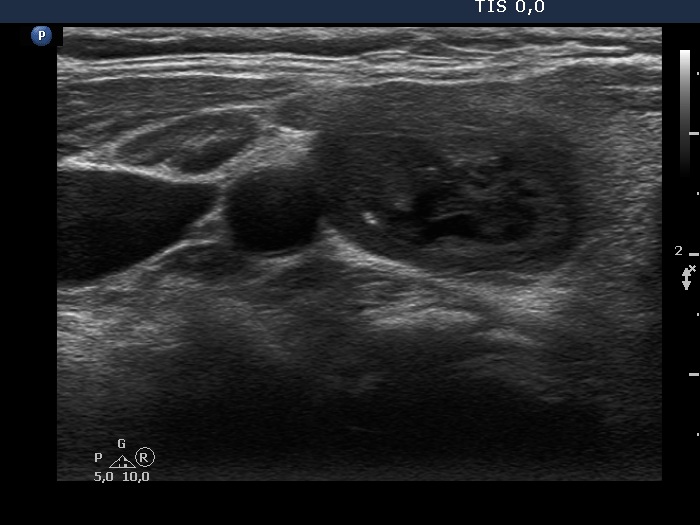
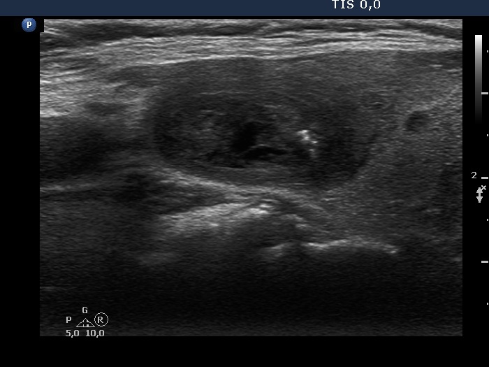
Ultrasonography. The thyroid was echonormal. There was a mixed moderately hypoechogenic-cystic nodule in the right lobe. The lesion presented various intranodular hyperechogenic granules including back wall figures, non-specific granules and a few typical comet-tail artifacts. There were other, difficult-to-classify bright granules. The latter could be either comet-tail artifacts or microcalcifications. There were several moderately hypoechogenic lesions in the left lobe.
3 mL brown fluid was aspirated. Cytology resulted in benign cystic degeneration.
Comments. This case illustrates the difficulty of distinguishing comet-tail artifact from microcalcification. The relevance of this differentiation lies in the consequences, the former is found almost always in benign lesions while the presence of microcalcifications increases the likelihood of papillary carcinoma.