
|
|
Intranodular hyperechogenic figures - case 402
|
|


Clinical data: A 28-year-old woman was referred for evaluation of elevated TSH level (5.94 mIU/L) detected on routine blood test. The patient's weight was 45 kg, her height was 167 cm.
Palpation: no abnormality.
Laboratory tests: TSH 6.06 mIU/L, FT4 17.1 pM/L, aTPO 0 U/mL, anti-hTg 1 U/mL.
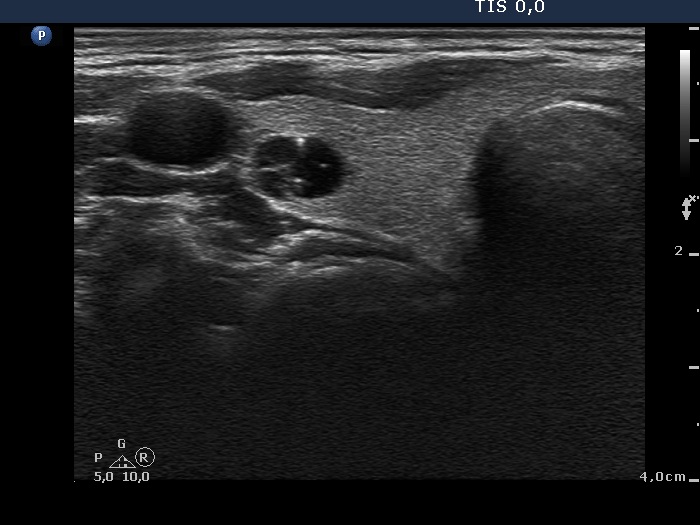
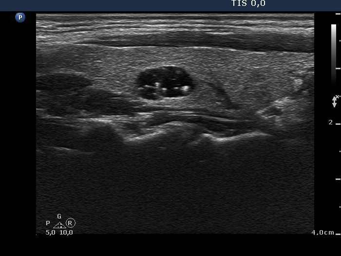
Ultrasonography. The thyroid was echonormal. There were several cystic lesions in both lobes which contained typical comet-tail artifacts. The nodule in the right lobe has back wall figures, too. The maximal diameter of the largest lesion was 11 mm.
Cytology resulted in colloid cyst.
We did not indicate replacement therapy because there were no signs of an underlying autoimmune thyroiditis and the patient has not yet planned pregnancy. A repeat TSH determination was advised in 6 months
Summary of follow-up. The elevated TSH level has spontaneously normalized and remained below 3 mIU/L in the next 3 years even during a pregnancy.
Comment. The normal value of the TSH is higher in thin patients.