Intranodular hyperechogenic figures - case 443 (ultrasonographic picture 13)
|
|

|
|
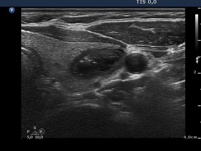
Left lobe, transverse scan - after aspiration of 2 mL brown fluid.

Echogenic figures
|
|
|
|
Left lobe, transverse scan - after aspiration of 2 mL brown fluid.