Intranodular hyperechogenic figures - case 662 (ultrasonographic picture 7)
|
|

|
|
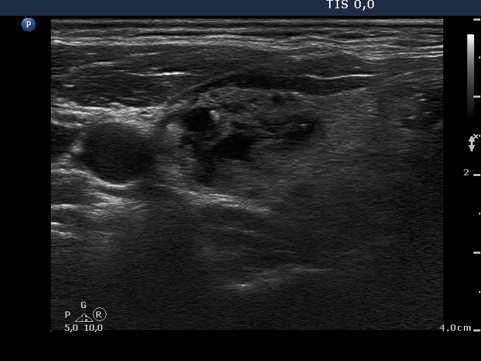
Right lobe, transverse scan - after aspirating 7.5 ml brown fluid. This is a multi-chambered, central-type cyst.

Echogenic figures
|
|
|
|
Right lobe, transverse scan - after aspirating 7.5 ml brown fluid. This is a multi-chambered, central-type cyst.