
|
|
Intranodular hyperechogenic figures - case 779
|
|


Clinical data: An 82-year-old woman was referred for evaluation of a thyroid nodule which was detected on physical examination. The patient noticed a lump in the left side of the neck 5 months ago. Otherwise, she had no complaints.
Palpation: a firm nodule in the right thyroid.
Functional state: euthyroidism with TSH 1.72 mIU/L.
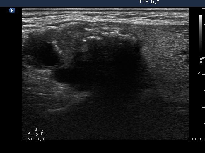
Ultrasonography. The thyroid was echonormal. There was a large cystic nodule in the upper pole of the left lobe. The lesion presented relatively large hyperechogenic granules. There were multiple enlarged lymph nodes lateral to the left lobe, the two largest were cystic.
US-guided aspiration was performed from the nodule. Cytology resulted in papillary carcinoma.
Total thyroidectomy was performed. Histopathology disclosed papillary carcinoma in the left lobe metastasizing to the lymph nodes.
Comment. The nodule in the left lobe had both macro- and microcalcifications. The latter can be observed in the form of clusters.