
|
|
Intranodular hyperechogenic figures - case 80
|
|


Clinical presentation: A 47-year-old woman was referred for evaluation of a lymph node in the neck and a nodular goiter. The problem was discovered during the examination of an upper airway infection.
Palpation: The right lobe was nodular on palpation. There was a painless node in the submental area.
Functional state: euthyroidism (TSH 1.82 mIU/L).
Ultrasonography: A large hyperechogenic nodule with coarse calcification occupied almost the entire right lobe. The nodule presented halo sign and scanty intranodular vascularity. There was a lymph node in the submental region. We did not find a regular hilum and the intranodal vascularization was increased.
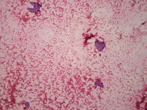
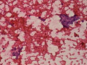
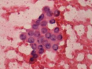
Cytological diagnosis of the thyroid nodule was suspicion of a Hürthle-cell tumor, while that of the lymph node was benign, reactive-type lymph node.
Wash-out thyroglobulin level of the lymph node resulted in 2.2 ng/mL while serum thyroglobulin was 24.7 ng/mL.
2 months later the patient underwent on right lobectomy. The submental lymph node was not found on preoperative neck ultrasound.
Histopathology disclosed benign hyperplastic nodules with oxyphilic metaplasia.
Comments.
-
The cytological pattern was identical with an oxyphilic tumor while the sonographic presentation stood against a follicular type tumor. Taking all these into account, an oxyphilic variant of papillary carcinoma had to be considered. However, there were neither inclusions nor grooves on the smear, therefore the most probable diagnosis was benign, hyperplastic nodule. Nevertheless, the possibility of an oxyphilic variant of papillary carcinoma could not be fully excluded.
-
The lack of regular hilum is an infrequent phenomenon in the case of a reactive-type lymph node, as was the irregular vascular pattern.