
Intranodular hyperechogenic figures - case 808 (ultrasonographic picture 1)

|
|
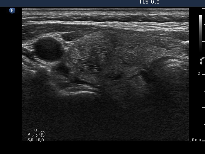
Right lobe, transverse scan. There are several hypoechogenic lesions in this section which contain pale granules and lines while the lateral one does more bight granules and lines, as well. The former figures correspond to normal while the latter to thickened connective tissue.