
|
|
Intranodular hyperechogenic figures - case 876
|
|


Clinical presentation: A 70-year-old woman has been followed for a thyroid nodule for 13 years. Cytology had resulted two-times in benign lesion. Two months before the present examination she was treated for hypertensive crisis in another hospital and she was told to harbor a very suspicious lesion in her thyroid which requires surgical removal irrespectively of cytological result.
Palpation: a not firm nodule in the right lobe.
Functional state: euthyroidism with TSH 1.37 mIU/L.
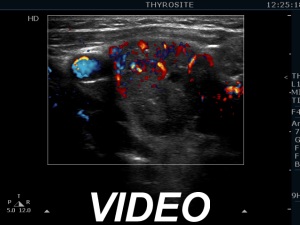
Ultrasonography. The thyroid was echonormal. There was a partially cystic nodule having an echonormal solid part in the right lobe. The lesion presented various hyperechogenic figures including comet-tail artifacts and ambiguous ones. The nodule did not grow in the past 13 years.
Aspiration cytology of the upper lesion resulted in benign cystic-colloid goiter.
Comments.
-
The nodule has equivocal echogenic granules. These are as bright as the comet-tail artifacts which are also present. It means that the ambiguous figures might be also colloid crystals though the possibility of punctate echogenic foci (microcalcifications) cannot be excluded.
-
This case does not belong to the exceptional cases in which we have to indicate surgery solely on the ultrasound presentation.