
Intranodular hyperechogenic figures - case 951
Follow-up examination 2 months later (ultrasonographic picture 2)
|
|

|
|
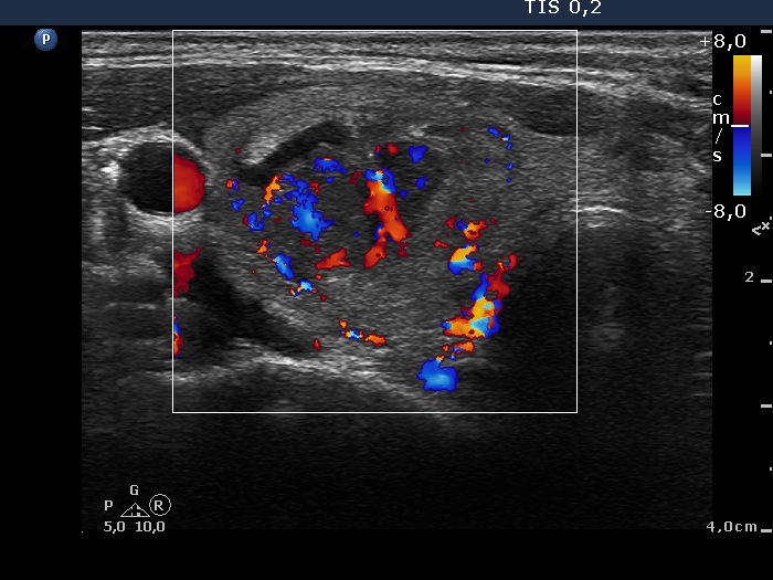
Upper part of the right lobe, transverse view, color Doppler mode. At this time, the nodule presents not only perilesional but even intralesional blood flow.